Testimonials
CT-FFR Assessment and Care Management
Corazon Imaging

Dr. John Rumberger and the team at Corazon Imaging have joined forces with Keya Medical to revolutionize the management of coronary artery disease through the power of AI CT FFR analysis.
FFRCT and DVFFR have transformed my practice, allowing me to provide superior patient care and achieve better outcomes. I am sincerely grateful for this invaluable resource provided by Corazon Imaging.
John A. Rumberger, PhD, MD, FACC, MSCCT, is a highly accomplished medical professional and currently holds the position of Medical Director at Corazon Imaging. His distinguished career includes serving as the Director of Cardiovascular Imaging at Princeton Longevity Center in New Jersey and as a former Professor of Medicine at the renowned Mayo Clinic. With his extensive fellowship in cardiology and an impressive portfolio of nearly 500 publications, he has made significant contributions to the field. Dr. Rumberger has also played a pivotal role in establishing the International Society of Atherosclerosis Imaging. Currently, he utilizes Keya Medical’s AI CT-FFR solution, DEEPVESSEL FFR, drawing upon his vast experience and expertise to enhance patient care at Corazon Imaging. He shares his experience with DEEPVESSEL FFR.
“I have recently gained access to FFRCT through my affiliation with Corazon Imaging. DVFRR, the FFRCT service provided by Keya, is not only reliable, but also user-friendly. The 3D model it offers has enhanced my understanding of stenosis and flow issues, allowing me to make more informed decisions.
One aspect that truly stands out is how DVFFR’s results have served as a crucial determining factor in cases where I faced uncertainty between medical therapy and follow-up catheterization. The accuracy and reliability of DVFFR’s results have been exceptional, giving me the confidence to make well-informed decisions.
The advanced AI capabilities of DVFFR have opened new horizons for me. By leveraging this technology, I have gained a deeper understanding of the vessel’s anatomy, surpassing what I could achieve solely through DICOM data. This enhanced comprehension has validated my initial suspicions about lesion significance and contributed significantly to my clinical experience and overall reporting confidence. Referring physicians have acknowledged and appreciated the added caution I can now provide.
I have integrated DVFFR into most cases where visual stenosis or consecutive stenoses measure 40% or higher, as it has become an indispensable tool for me. Additionally, I have carefully utilized DVFFR to interpret flow patterns in patients with previous stents, relying on my extensive experience in clinical imaging. This approach allows me to assess the potential for distal ischemia, ensuring comprehensive patient care.
FFRCT and DVFFR have transformed my practice, allowing me to provide superior patient care and achieve better outcomes. I am sincerely grateful for this invaluable resource provided by Corazon Imaging. It has undoubtedly elevated my expertise and brought immense value to my clinical practice.”
Case Study
AI-based CT-FFR Assessment and Management of Multivessel Coronary Artery Disease with High-Risk Plaques
Abstract:
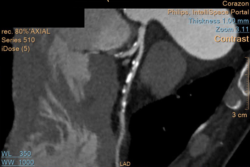
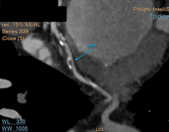
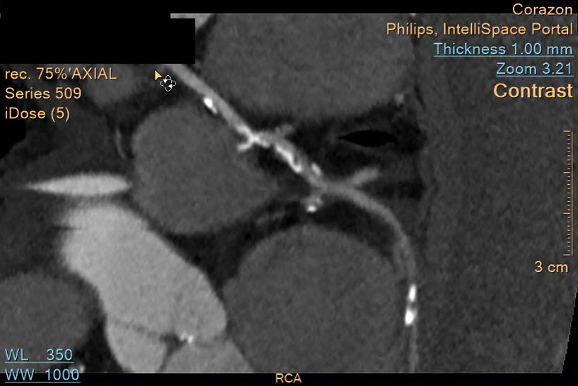
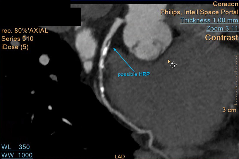
This case study presents a 56-year-old female patient with palpitations, shortness of breath with effort, hypertension, high cholesterol, diabetes, and a family history of coronary artery disease (CAD). Contrast-enhanced CT imaging revealed extensive coronary calcifications, moderate stenoses, and high-risk plaques in the LAD and LCX. DEEPVESSEL FFR CT analysis confirmed multi-vessel obstructive CAD. The patient was prescribed Rosuvastatin, Ezetimibe, and aspirin and underwent cardiac catheterization to assist with revascularization alternatives. The study emphasizes the importance of comprehensive diagnostics, including AI-driven FFR analysis, for identifying and managing multivessel CAD with high-risk plaques.
Introduction:
Coronary artery disease (CAD) is a leading cause of morbidity and mortality. Timely diagnosis and treatment are critical. This case study discusses the diagnostic process, risk assessment, and treatment strategy for a patient with multivessel CAD and high-risk plaques, utilizing DEEPVESSEL FFR CT analysis.
Case Presentation:
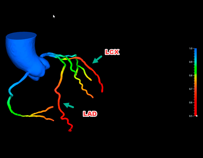
A 56-year-old female patient with shortness of breath with effort, palpitations, and cardiovascular risk factors underwent contrast-enhanced CT imaging. Extensive coronary calcifications, moderate stenoses, and high-risk plaques were detected in the LAD and LCX. DEEPVESSEL FFR CT analysis confirmed the hemodynamic significance of the lesions (LAD: .73, LCX: .66, RCA: .75).
Diagnostic Approach:
Contrast-enhanced CT imaging with a Philips 128-slice MDCT Scanner provided detailed anatomical information, identifying calcifications, stenoses, and high-risk plaques. DEEPVESSEL FFR CT analysis assessed the hemodynamic significance of the lesions in the LAD, LCX, and RCA.
Management and Treatment:
Due to the indication of double-vessel obstructive CAD with high-risk plaques, the patient underwent cardiac catheterization. Rosuvastatin, Ezetimibe (10 mg/day), and aspirin were also prescribed to manage cholesterol levels and reduce thrombotic events.
Conclusion:
This case underscores the significance of a comprehensive diagnostic approach, including AI-driven FFR analysis, in a patient with dyspnea, palpitations, and cardiovascular risk factors. Contrast-enhanced CT imaging and DEEPVESSEL FFR CT analysis aided in identifying extensive calcifications, stenoses, high-risk plaques, and at least double vessel obstructive CAD.
Clemenceau Medical Center (Johns Hopkins Medicine International)
Clemenceau Medical Center in Beirut, Lebanon, is a leading hospital affiliated with Johns Hopkins Medicine International. Known for its state-of-the-art facilities and cutting-edge medical services, CMC offers a wide range of specialties, including cardiovascular medicine, oncology, orthopedics, and neurology. The center is recognized for providing high-quality patient care and advanced diagnostic technologies. Additionally, CMC emphasizes medical education, research, and international collaborations.
Clemenceau Medical Center implements cardiovascular diagnostics CT FFR with DeepVessel FFR.
With CT scanning, blockages in the coronary arteries can be detected, and FFR CT takes it further by measuring blood flow past the blockages at multiple points along the artery. Unlike traditional invasive FFR, which only gives a reading at a single segment, this advanced technology is now available at CMC, making it the first in Lebanon and the Middle East to offer care comparable to Europe and North America.

