CCTA Acquisition Requirements
Compliance with Coronary Computer Tomography Angiography (CCTA) technical specifications.
1. A high-resolution CT machine must acquire images with at least 64 rows.
2. The imaging area should encompass all coronary arteries and the adjacent ascending aorta.
3. The reconstructed CT layers should be free from any abnormalities.
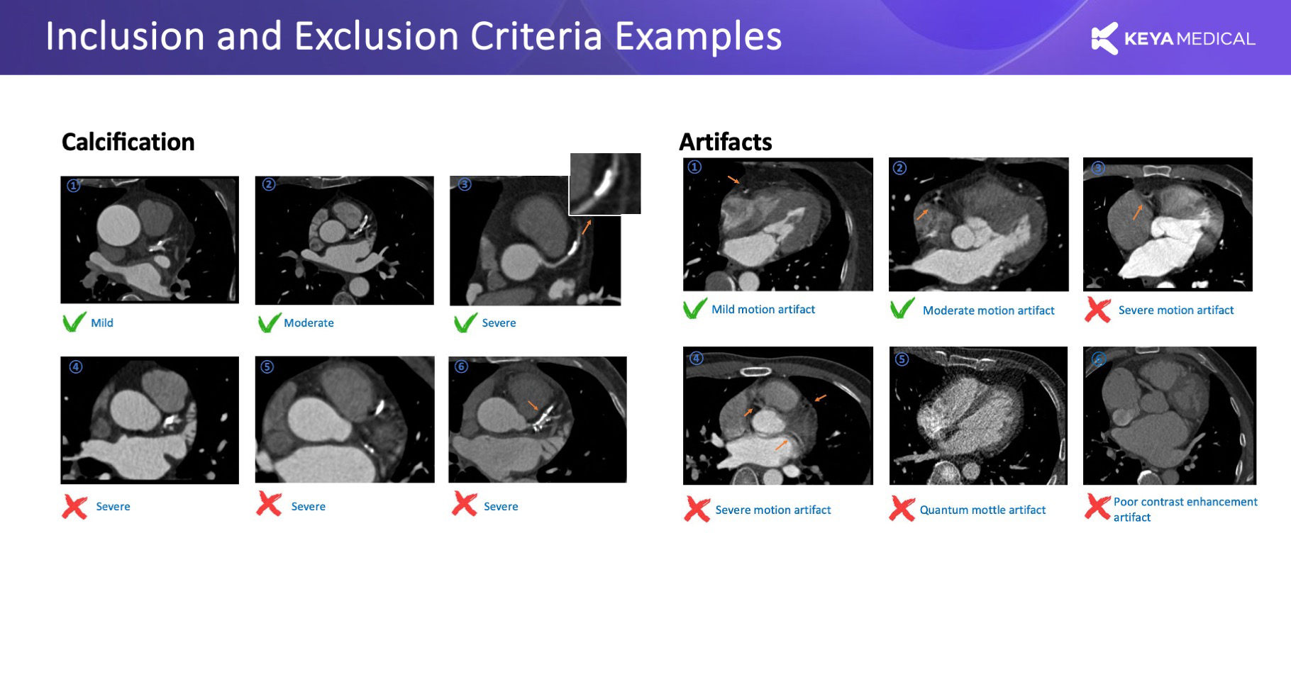
4. In 3D reconstruction, no affected area should exceed a diameter of 2cm due to factors such as motion artifacts, image noise, or vessel contrast ratios.
5. Patients with metallic stent or bypass grafting in any coronary artery should be excluded.
6. CT Key Parameter Requirements:
- Layer thickness ≤1mm
- In-plane interval ≤0.5mm
- Layer spacing ≤1mm.
- KVP≥70
7. Heart Rate Control:
- For CT machines with 64 rows, maintain the patient’s heart rate below 70 bpm.
- For CT machines with 64+ rows, maintain the patient’s heart rate below 90 bpm.
8. Coronary vasodilation
- Give 0.4-0.8 mg sublingual nitroglycerin (tabs or spray) 5 min before contrast.
- Omit nitroglycerin in patients with contraindications (i.e., aortic stenosis, recent intake of phosphodiesterase inhibitors, etc.).
Image Quality Assessment - Likert Scale Rating and Examples
CCTA images must meet high-definition criteria and be acquired using a CT scanner with 64 slices or more. The imaging field should encompass all coronary arteries and the adjacent ascending aorta. It is crucial to prevent image motion artifacts, as they can interfere with the interpretation and 3D reconstruction of blood vessels larger than 2mm in diameter, alongside issues related to noise, contrast agent concentration, and other factors.
Data associated with non-indications such as implantable defibrillators (ICDs), pacemakers, coronary artery bypass grafting (CABG), valve replacement surgery, cardiomyopathy, etc., must be carefully interpreted and excluded from the analysis.
5-Point Image Quality Evaluation Form
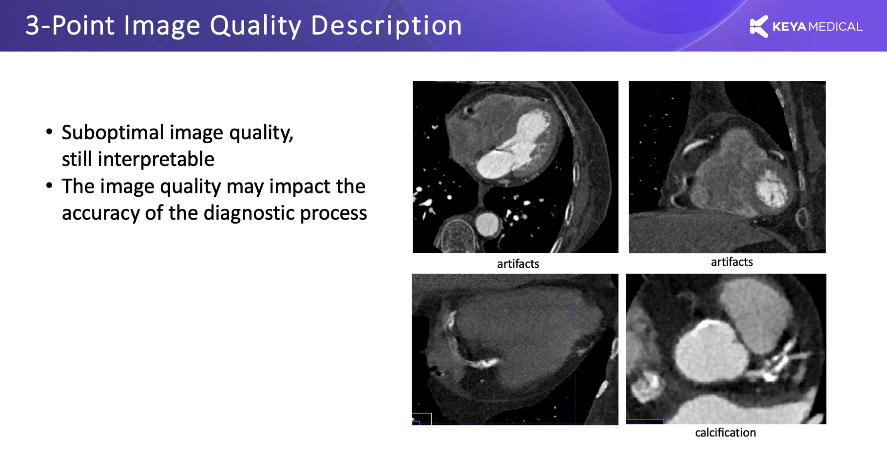
Images with 3 points or higher meet the diagnostic quality requirements.
| Scale | Description |
| 5-point | Excellent image clarity, distinct vessel boundaries, and flawless display with zero motion artifacts. |
| 4–point | Good quality imaging, generally clear boundaries, minimal motion artifacts that have negligible impact on the diagnostic process. |
| 3–point | Fair image quality, is still interpretable for diagnosis, though there may be some influence on the accuracy of the diagnosis. |
| 2–point | Poor image quality, is still interpretable for diagnosis, though there may be some influence on the accuracy of the diagnosis. |
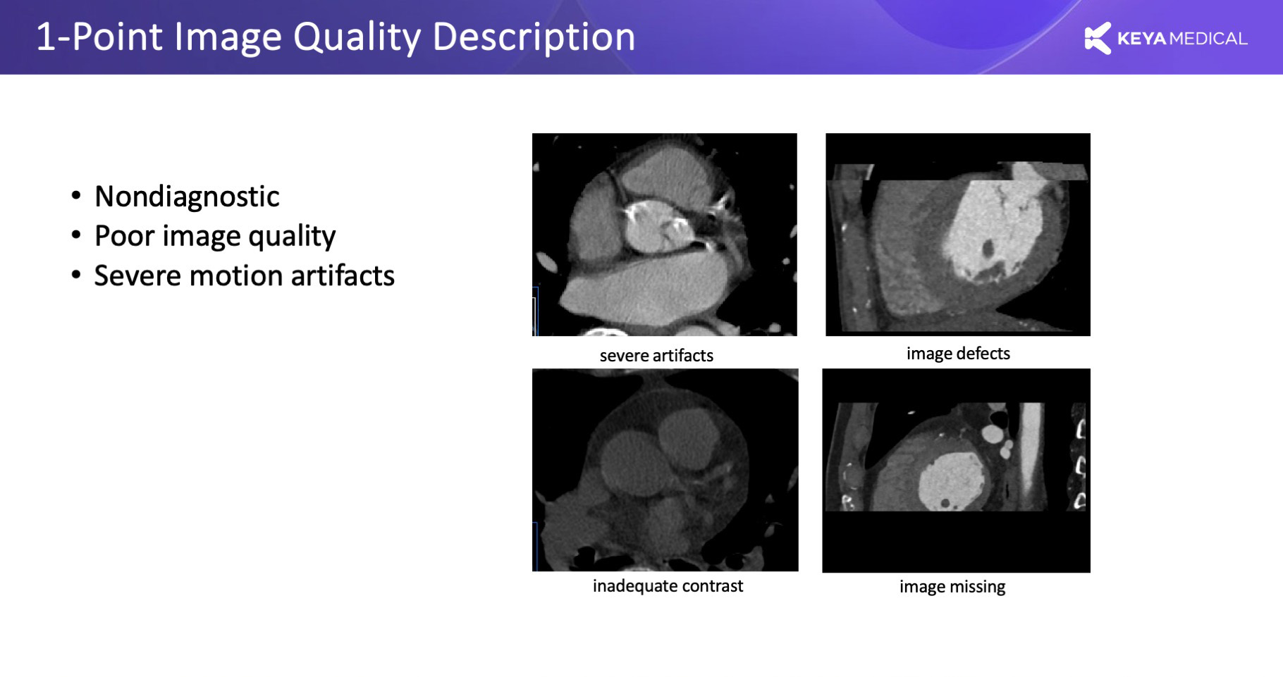
| 1–point | Non-diagnostic images are characterized by poor quality and severe motion artifacts. |


